A clubfoot is usually visible at birth, but its effects extend beyond appearance: without correction, the foot can remain stiff, poorly aligned and unable to provide a normal surface for walking. By understanding the warning signs, treatment timeline, casting and bracing phases, parents can seek assessment early and support a more functional result.
Key takeaways
- Have a newborn assessed promptly when the foot turns inward or points downward.
- Ponseti treatment gradually corrects cavus, adduction, varus and equinus.
- Use the prescribed brace after correction to reduce relapse risk.
- Keep follow-up visits to monitor growth, alignment and long-term foot function.
How untreated clubfoot can affect walking and foot development
Untreated clubfoot changes both shape and mechanics. A high arch (cavus) lifts the inner midfoot, the forefoot points inward (adduction), the heel turns inward (varus), and the ankle points downward (equinus). These clubfoot symptoms at birth are not a temporary position that reliably straightens.
| Feature | Untreated clubfoot | After early correction |
|---|---|---|
| Weight-bearing surface | Rigid and angled, rather than plantigrade | More evenly aligned for standing and walking |
| Pressure pattern | Outer-edge loading can cause calluses and painful pressure areas | Weight spreads across more of the sole |
| Movement | Abnormal gait, difficulty with balance and push-off | Better support for walking, running and play |
| Footwear | Ordinary shoes may not fit comfortably | Shoes usually fit more easily |
As the child grows, the stiff foot cannot develop a normal plantigrade weight-bearing surface. In clubfoot and walking, this can mean walking on the outside border, an abnormal gait, painful rubbing, difficulty wearing ordinary shoes and reduced activity.
Early correction gives the developing foot a better platform for weight-bearing and helps prevent deformity from becoming harder to change. This is why timing matters in clubfoot foot development.
Correction does not make the feet identical. The treated foot may remain smaller, the calf thinner, and ankle or subtalar motion reduced; many children still walk, run and participate normally.
When a newborn should be assessed and how clinicians confirm clubfoot
Arrange a clubfoot assessment during the first weeks after birth, even though treatment is not usually an emergency on the day of delivery. Early referral lets the clinician plan Ponseti care while the soft tissues remain adaptable; waiting can make correction more demanding.
Clubfoot diagnosis usually comes from the newborn examination, not a scan. The clinician checks:
- The foot’s inward and downward shape, high arch, heel position and skin creases
- Flexibility and the ability to move the foot
- Pulses, muscle activity, toe movement and skin condition
- Whether the foot is typical, rigid, difficult to classify or linked to another physical finding
A typical isolated clubfoot does not need routine imaging. A clubfoot X-ray or other testing becomes useful when the deformity is atypical, unusually stiff, uncertain after examination or associated with another condition.
The assessment must also consider arthrogryposis, spina bifida, and neuromuscular or genetic disorders because associated clubfeet are often stiffer and require a broader treatment plan.
For families in Nagpur, Dr. Girish Motwani Best Orthopedic Surgeon - SUYOG can provide an orthopedic assessment and help plan referral when the diagnosis or associated findings are unclear. Ask specifically when to start clubfoot treatment, whether the foot is idiopathic or associated, and which examination findings will determine the next step.
Related product
 | Congenital foot anomaly like club foot,vertical talus,Hemimelia Congenital foot anomaly like club foot,vertical talus,Hemimelia Congenital foot anomaly like club foot,vertical talus,Hemimelia View product → |
What early Ponseti treatment does to the developing foot
The Ponseti method reshapes an idiopathic clubfoot gradually, using gentle manipulation followed by long-leg casts rather than forceful one-time correction. The sequence matters because correcting the parts in the wrong order can create a residual or rocker-bottom deformity.
| Component | Correction sequence | What to avoid |
|---|---|---|
| Cavus | Lift the first metatarsal to lower the high arch | Pressing the forefoot downward |
| Forefoot adduction | Turn the forefoot outward while supporting the talar head | Pronating the forefoot |
| Hindfoot varus | Bring the heel into alignment as the forefoot corrects | Trying to correct every component at once |
| Ankle equinus | Correct the downward-pointing ankle last | Forcing ankle dorsiflexion before the heel and forefoot align |
Expect several casts, commonly five to seven, changed at roughly weekly intervals. After each cast, check the toes for normal color, warmth and movement, and look for swelling or skin irritation. Keep the cast dry and intact; never push a stick, cotton swab or other object inside it to scratch.
Persistent equinus after casting is commonly treated with a percutaneous Achilles tenotomy. This brief planned step is part of standard Ponseti care, not proof that casting failed.
Early treatment uses the newborn’s more moldable soft tissues, so extensive early surgery is not the modern default for idiopathic clubfoot; large releases can leave stiffness, weakness and later pain.
Why bracing matters after the foot has been corrected
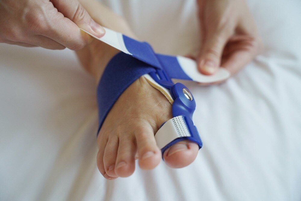
Casting creates the correction; the clubfoot brace holds each foot in its prescribed position while the muscles, ligaments and joints adapt. Without that support, the corrected foot can gradually turn inward again during early childhood.
A commonly used schedule is near-full-time wear for about three months, followed by nighttime and nap-time use through approximately age four or five. This pattern is not universal, so follow the treating clinician’s schedule and the device manufacturer’s fitting instructions. Age, severity, associated conditions and recurrence history can all change the plan.
Clubfoot brace adherence is one of the strongest modifiable influences on relapse, making the foot-abduction brace central to clubfoot relapse prevention. A child with an excellent initial correction can develop renewed inward turning if bracing stops early or becomes inconsistent.
Contact the clinic for adjustment if you notice:
- The heel slipping out of the shoe
- Red marks that do not fade after the brace is removed
- Blisters or broken skin
- Heavy sweating or difficulty tolerating the brace
Do not abandon the brace because it seems uncomfortable. An incorrect fit, a growing foot or a strap problem often explains the difficulty, and prompt adjustment can protect the correction.
How follow-up protects the child’s long-term foot function
Follow-up protects long-term foot function after both casting and bracing. At visits, the clinician checks ankle dorsiflexion, foot alignment, muscle balance, skin, brace fit and how the child walks. Clubfoot relapse signs include reduced ankle dorsiflexion, renewed inward turning or walking on the outside border of the foot.
Report these changes promptly. Early detection gives the clinician more options before the foot becomes stiff. Recurrence does not automatically mean permanent damage or extensive surgery: renewed casting, repeat Achilles lengthening or another limited procedure may correct an early relapse, while a late rigid relapse can require more extensive treatment.
Brace adherence remains one of the strongest modifiable influences on relapse. Contact the clinic about a slipping heel, persistent red marks, blisters, sweating or intolerance instead of stopping the brace. The goal is a comfortable, plantigrade foot that fits shoes and supports walking, running and play—not feet that look identical.
A treated foot may remain smaller, with a thinner calf or reduced ankle and subtalar motion.
An associated clubfoot needs broader evaluation and a different plan than idiopathic clubfoot, especially when arthrogryposis, spina bifida or a neuromuscular or genetic condition is present. Ask:
- Is the foot idiopathic or associated with another condition?
- Is correction progressing in the expected order?
- When can the brace schedule change?
- What signs require an earlier review?
Frequently asked questions
How can untreated clubfoot affect walking and foot development?
Untreated clubfoot can leave the foot curved inward and pointed downward, making walking difficult and causing abnormal weight-bearing, shoe problems and reduced foot function.
When should a newborn be assessed for clubfoot?
Arrange an assessment soon after birth when the foot has a high arch, inward-pointing forefoot, inward-turned heel or downward-pointing ankle.
How do clinicians confirm clubfoot?
Clinicians examine the foot’s shape, flexibility and alignment, then distinguish true structural clubfoot from a flexible positional deformity. Imaging is not always needed.
What does early Ponseti treatment do to the developing foot?
Ponseti treatment uses gentle manipulation and a sequence of casts to gradually correct the foot’s cavus, adduction, varus and equinus.
Why is bracing important after clubfoot correction?
The brace holds the corrected position while the child’s tissues develop, helping prevent the foot from gradually returning to its clubfoot position.