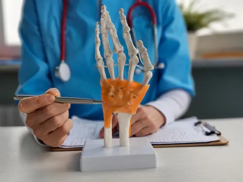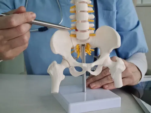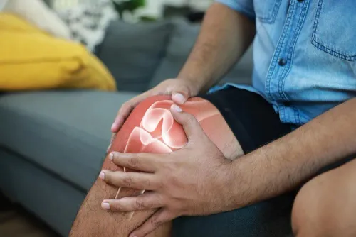
A damaged joint is not automatically a lost joint, but preservation depends on what caused the damage, how much tissue remains, and whether the joint still has a workable mechanical foundation. By the end, you will know which findings support preservation, which treatments address the source of damage, and when replacement or fusion becomes the more realistic option.
Key takeaways
- Preservation targets a repairable cause, not diffuse bone-on-bone arthritis.
- Alignment, stability, cartilage loss, and symptoms determine candidacy.
- Ask how imaging findings connect to the proposed repair.
- Replacement becomes more suitable when damage is widespread or preservation cannot restore function.
What Does Joint Preservation Actually Mean?
Joint preservation means keeping your natural joint functional by correcting the cause of damage, such as instability, poor alignment, a focal cartilage defect, a loose body, or a torn ligament. It does not mean regrowing an entire joint or reversing diffuse, bone-on-bone arthritis.
The aim is to delay or avoid joint replacement or joint fusion while retaining useful movement.
| Option | What it means | How it differs from joint preservation |
|---|---|---|
| Joint preservation | Repairs or corrects a specific problem in the existing joint | Addresses mechanics or focal damage rather than replacing the joint |
| Joint replacement | Removes damaged joint surfaces and inserts an implant | Replaces the joint when damage is too widespread for repair |
| Joint fusion | Permanently joins the bones | Relieves pain by eliminating movement at that joint |
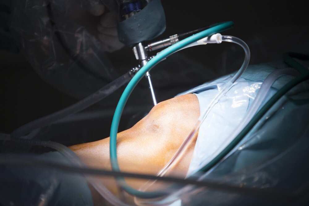
| Arthroscopy | Uses a camera and small instruments through tiny incisions | A surgical method that can support preservation, but is not itself a treatment goal |
| Injections | Places medicine such as corticosteroid or hyaluronic acid into the joint | Can reduce symptoms temporarily but does not correct deformity or restore damaged cartilage |
| Bracing | Supports alignment or limits painful movement from outside the body | Controls load while the joint remains unchanged |
| Physiotherapy | Builds strength, mobility, balance, and movement control | Improves function without directly repairing structural damage |
| Observation | Tracks symptoms, examination findings, and imaging over time | Avoids intervention while damage and function remain acceptable |
Preservation surgery can still involve months of protected weight-bearing and rehabilitation. Keeping the joint is not the same as guaranteeing normal movement or immediate pain relief.
Which Damaged Joints Are Candidates for Preservation?
A focal cartilage defect, a torn ligament, or abnormal alignment can make preservation realistic; diffuse bone-on-bone arthritis usually does not. The key question is whether a correctable problem remains, rather than whether the joint looks damaged.
Your patient factors matter as much as the scan:
- Patient age and activity level shape the trade-off between recovery time, implant longevity, and return to impact sports or heavy work.
- Pain pattern matters: pain linked to one loading position suggests a focal problem, while constant rest or night pain points toward advanced joint disease.
- Joint stability, range of motion, walking tolerance, and functional limitation show whether the joint still works after the painful area is addressed.
- Your willingness to follow protected weight-bearing and rehabilitation can determine whether repair or realignment succeeds.
Joint factors include cartilage loss, alignment, deformity, bone quality, and the cause of damage. A stable joint with preserved cartilage around a local defect offers a different opportunity from a deformed joint with widespread cartilage loss.
| Finding | Preservation is more realistic when | Another plan becomes more likely when |
|---|---|---|
| Focal damage | One cartilage or osteochondral defect has healthy surrounding joint surfaces | Cartilage loss is widespread |
| Mechanical problem | Alignment or ligament failure is correctable | Severe deformity cannot be corrected safely |
| Bone and soft tissue | Bone, blood supply, and skin can support healing | Major bone loss, avascular necrosis, infection, or poor tissue quality is present |
| Disease pattern | Injury or wear affects one area | Inflammatory arthritis or end-stage arthritis affects the whole joint |
Even a technically preservable joint may not justify months of restricted activity if pain is modest and function remains good. Conversely, severe pain with limited X-ray changes deserves assessment because symptoms and visible damage do not always match.
How Do Clinicians Measure the Damage?
A joint is not judged by one scan or by pain alone. Clinicians combine your pain pattern, walking ability, range of motion, tenderness, stability, strength, limb alignment, and activity goals with the images. This matters because severe pain can occur with modest X-ray changes, while extensive joint-space loss may cause surprisingly little disability.
Each assessment answers a different question:
- Examination shows whether instability, restricted motion, tendon weakness, altered gait, or a tender focal defect is driving the damage. It also reveals whether the joint remains usable under everyday load.
- Weight-bearing X-rays show joint-space loss, bone spurs, deformity, and alignment while you stand. Alignment views clarify whether abnormal loading is concentrated on one part of the joint and whether an osteotomy could redirect force.
- An MRI cartilage scan shows cartilage thickness and defects, bone marrow changes, osteochondral lesions, tendons, ligaments, and hidden damage that an X-ray can miss. It is not automatically needed for every case of suspected arthritis.
- A CT scan creates detailed cross-sectional and three-dimensional views of bone. It helps define cysts, loose fragments, fractures, bone loss, and complex deformity before reconstruction or corrective surgery.
A focal cartilage defect with preserved alignment and stability is different from diffuse bone-on-bone arthritis. The findings also indicate whether cartilage treatment must be combined with ligament reconstruction, tendon balancing, or correction of the load-bearing axis; treating the visible defect alone can leave the damaging force in place.
Which Treatments Repair the Cause Instead of Only Easing Pain?
Activity modification, weight management, physiotherapy, and anti-inflammatory medicines are chosen when symptoms arise from overload, weakness, stiffness, or early irritation rather than a structural defect needing surgery. They reduce the damaging load and restore movement, but they cannot reattach a torn ligament or replace missing cartilage.
| Option | What it repairs or changes | When it applies |
|---|---|---|
| Non-surgical care | Load, strength, flexibility, and inflammation | Pain without a repairable structural lesion, or as preparation for surgery |
| Cartilage procedure | A local cartilage or osteochondral defect | A contained defect with preserved surrounding joint, suitable alignment, and limited arthritis |
| Tendon or ligament repair | A torn or elongated stabilising structure | Instability or loss of tendon function causing abnormal joint loading |
| Osteotomy | The bone’s load-bearing axis | Malalignment concentrates force on one compartment or threatens a cartilage repair |
Cartilage treatment is not a cure for broad bone-on-bone arthritis. Microfracture creates fibrocartilage rather than normal hyaline cartilage; graft transplantation and autologous chondrocyte implantation suit different defect sizes and bone conditions. If the ankle is unstable or misaligned, the surgeon may combine cartilage work with ligament reconstruction, tendon balancing, or osteotomy.
Every surgical option carries a recovery cost: protected or non-weight-bearing time followed by structured rehabilitation, with higher-impact activity delayed for months. Smoking or nicotine exposure can impair bone healing, so cessation is especially important before osteotomy. The choice must match the defect, its cause, and your willingness to complete rehabilitation.
When Does Preservation Stop Being the Right Goal?
Preservation stops being the right goal when damage is widespread rather than confined to a repairable area. Bone-on-bone cartilage loss, major bone loss, severe deformity, infection, inflammatory arthritis, avascular necrosis, or an unstable joint can make repair unlikely to deliver lasting relief.
The next option depends on the joint, the scan findings, and what you need from it:
- Total ankle replacement suits selected people with end-stage ankle arthritis who want to retain some ankle motion and have adequate bone, blood supply, and soft-tissue cover. Severe deformity, neuropathy, active infection, poor bone quality, or high-impact activity can raise loosening and revision risks.
- Ankle fusion suits advanced arthritis when reliable pain relief matters more than ankle movement, or when replacement is unsafe. It removes ankle motion, which can make uneven ground and stairs harder and increase demands on nearby foot joints; failure of the bones to unite is another risk.
- Specialist assessment is essential when a focal osteochondral talar defect is being considered for cartilage restoration. Grafting or marrow-stimulation treats a defined cartilage-and-bone problem, not diffuse arthritis, and lesion size, alignment, bone involvement, and rehabilitation demands affect the choice.
Your priorities matter too. A person who needs heavy-impact work may prefer a durable pain-relieving option, while someone valuing motion may accept longer rehabilitation and implant surveillance. Dr. Girish Motwani Best Orthopedic Surgeon - SUYOG can help match ankle imaging, activity demands, and risks before choosing preservation, replacement, or fusion.
Related items
 | Osteochondral defects of Talus Osteochondral defects of Talus Osteochondral defects of Talus View product → |
Orthopedic Doctors Orthopaedic all orthopedic services available |
Frequently asked questions
What does joint preservation mean?
Joint preservation keeps your natural joint functional by correcting a specific cause, such as instability, poor alignment, a focal cartilage defect, a loose body, or a torn ligament. It does not regrow an entire joint or reverse diffuse bone-on-bone arthritis.
Which damaged joints are candidates for preservation?
Candidates include joints with a local cartilage defect, ligament tear, loose body, or correctable alignment problem. Clinicians also consider your symptoms, activity limits, joint stability, and the extent of damage on imaging.
How do clinicians measure joint damage?
They combine your symptom history and physical examination with imaging. X-rays show alignment and joint-space loss, while MRI can show cartilage, ligaments, menisci, bone marrow changes, and loose bodies.
Which treatments repair the cause of joint damage?
Treatment depends on the cause and may include ligament repair or reconstruction, removal of a loose body, cartilage procedures, or realignment surgery. The aim is to address the structural problem rather than only reduce pain.
When does joint preservation stop being the right goal?
Preservation becomes less suitable when cartilage loss is widespread, the joint is bone-on-bone, deformity is severe, pain and disability persist despite treatment, or a repair cannot restore useful function. Joint replacement may then offer a more reliable solution.
Related items
 | Achilles Tendon Injuries Treatment Achilles Tendon Injuries Treatment Achilles Tendon Injuries Treatment View product → |
 | Ankle and foot arthritis Ankle and foot arthritis View product → |
 | Congenital foot anomaly like club foot,vertical talus,Hemimelia Congenital foot anomaly like club foot,vertical talus,Hemimelia Congenital foot anomaly like club foot,vertical talus,Hemimelia View product → |
Orthopedic all xray View service → |